Carcinoid Triad

Another kind of Triad: The Three Body Problem

Carcinoid Tumors and Anesthesia: Notes from TrueLearn

Carcinoid Tumors: Slow growing, benign, small intestine tumors that can metastasize

Hormonal secreting tumors -> cause cutaneous flushing of head neck and thorax, bronchoconstriction, hypotenion, diarrhea, heart disease

Carcinoid Crisis: triggered by physical/chemical such as histamin release, serotonin, bradykinin

Other triggers include: chemo, tumor necrosis, or succinycholine induced fasciculations!

Anesthestic management: avoid histamine release (succ, atracurium, thiopental, morphine, vancomycin); Desflurane good for patients with liver metastasis as low hepatic metabolism (0.02%); Also the use of NE, epi, Dopamine and isoproternol a/w carcinoid crisis

Carcinoid Heart Disease seen in 60% of patients with carcinoid ->right side:  tricuspid and pulmonic valves (plaque like deposits on valves) with TR as most common finding; 50% of carcinoid deaths are from cardiac involvement

So Carcinoid Triad = flushing, diarrhea and cardiac involvement

Management I/O And Peri-Op:

Carcinoid tumors secrete variety of substances (serotonin, catecholamines, histamine)

Somatostatin therapy is standard of care

Be prepared for rapid BP changes
-alpha/beta blockers for HTN
-Vaso for hypotension, or neo

Avoid: Beta agonists -> increased release of from carinoid !

https://www.openanesthesia.org/carcinoid_crisis_treatment/


Levo vs. Neo: A Change of the Guards?

Vancomycin IV infiltration, yet we still give through PIV

From A&A May 2016: Should Norepinephrine, Rather than Phenylephrine, Be Considered the Primary Vasopressor in Anesthetic Practice?

http://www.ncbi.nlm.nih.gov/pubmed/27101504

GA = Sympathectomy, decreased NE and Epi concentrations

Phenylephrine (PE, trade name Neosynephrine aka Neo)
pure alpha 1 agonist
increase ABP, decrease HR
CO effect mixed +/- venous return, decreased HR

Norepinephrine (NE, trade name Levophed aka Levo)
alpha1 and beta1, some beta2
relaxation of venous resistance
NE enhances venous return +CO
+ionotropy, little chronotropy
+arterial vasoconstrict
+HR, +SV, +CO

Potency of NE:PE 20:1

NE levels increased in cardiac failure

PeriOp circulating Catecholamine levels

NE and Epi decreased by
-diazepam
-thiopental (even more in uremic)
-midazolam
-pancuronium
-spine surgery + prop or iso

NE and Epi increased by
-Ketamine
-1.5 mac Desflurane
-succinylcholine
-rapid increase in Isoflurane
-intubation
-abdominal surg + sevo/n2o
-emergence/extubation

little effect from roc/vec

Comparing NE to PE in specific studies and circumstances

Aortic stenosis
-PE resulted in higher MAP, no differencve in LV fx vs. Levo

CABG
-PE imparied LV function, unchanged with levo

Pulmonary HTN cardiac surgery pts
-CI maintained with NE, decreased with PE
-NE improved (reduvced) ratio of mean PA pressure to SBP

General Surgery, isoflurance
-NE increased LV performance
-PE decreased LV performance

General Surgery, prop+remi
-both PE/NE decreased CO and HR, increased MAP

C/S Spinal
-PE vs. NE infusion
-NE had same sbp, higher HR, higher CO c/w PE

If LV fx impaired, enhanced afterload from PE -> decreased CO and systemic perfusion

but what about infiltration? Largely overblown as, yes NE 7x more constrictive in radial ARTERY, yet only 76% more vasoconstrictive in veins and concentration of Levo is 20x less than PE

if infiltrate -> give phentolamine


Coronaries: Considerations

coronary (adj.)Look up coronary at Dictionary.com
c. 1600, “suitable for garlands,” from Latin coronarius “of a crown,” from corona “crown” (see crown (n.)). Anatomical use is 1670s for structure of blood vessels that surround the heart like a crown. Short for coronary thrombosis it dates from 1955. Coronary artery is recorded from 1741.

Anesthetic Considerations for Coronary Artery Disease patients, from Open Anesthesia

General:

  • avoid swings: keep normotensive (20% baseline) and baseline HR
  • Closely watch I/O EKG as 1mm ST change (+/-) for >60s = 10x cardiac event risk
  • 5 mins of hr >105 = 10x death risk p/o

Imaging/Studies

  • Assess echo, stress tests, viability studies, cath for LV fx, valve abnormalities, CAD
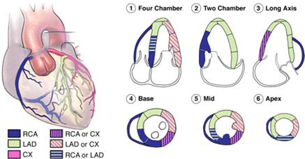
  • Quick Guide images: 
    Distribution of Coronary Arteries in relation to Echo Segments
    Distribution of Coronary Arteries in relation to Echo Segments


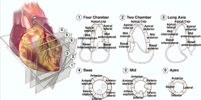
    echo sections
    Echo Segments and 3D View


Starting Off: Induce and Intubate

  • Etomidate or low dose propofol, Fentanyl + Midazolam
  • DL < 15s (be quick!)
  • if anticipate difficult intubation, consider spraying lidocaine, blocks, or iv lido prior to DL

Keep it going: Maintenance and Monitoring

  • Debate between Volatiles vs. N20 Opiate, conflicting data, use judgement
  • Volatiles depress but also provide preconditioning that is helpful
  • Short acting BB (Esmolol) not proven to help, but use judgement
  • Paralytics: Pancuronium increases BP and HR, Atracurium and miv can lower BP, Vec/Reoc/Cis neutral (though some concern about histamine with Cis, much lower than Atracurium)
  • Don’t reverse: glyco not well tolerated
  • Standard cardiac setup if high risk surgery (cardiac) or high risk pt (recent MI, CHF, unstable angina)
  • NTG good for ischemia

Finish up: Avoid hypothermia, adequate analgesia and sedation en route to ICU


Who is Brugada?


There is not just one Brugada, but a family of them!

It all began in 1987 when Professor Brugada was running the electrophysiology laboratory at the University of Maastricht in the Netherlands and a Polish man brought his son to see him. The boy was 3 years old and had a history of repeated episodes of fainting and cardiac arrest; his father had resuscitated him several times. His sister had suffered similar symptoms and had died at the age of 3 in spite of treatment with a pacemaker and amiodarone.
“The electrocardiogram of that boy was never seen before and something you could not find in any publication,” Professor Brugada remembers. “I was very fortunate that the father was able to return to Poland and bring the electrocardiograms of the sister, which turned out to be exactly the same as those of the brother. Remember, it was 1987; the Berlin Wall was still there, so everything that this guy was doing was illegal.”
It took 4 years for the Brugadas to find 2 more patients with similar ECGs – one from the Netherlands and the other from Belgium. They presented the 4 cases as an abstract to an American Heart Association conference, and several doctors who had come across similar cases contacted them. A year later, they published details of 8 patients [in JACC: http://www.sciencedirect.com/science/article/pii/073510979290253J%5D.

AICD Indications

Secondary Prevention of death due to VF/VT in settings of:

1. prior VT/VF req’ing resuscitation, or unstable VT with unknown cause
-includes idiopathic vf/vt and congenital long qt
-excludes vt/vf w/in 48hrs of MI

2. spontaneous sustained VT in presence of heart disease (valvular, ischemic, hypertrophic, dilated, infiltrative cardiomyopathy or channelopathies)

Primary Prevention

For the primary prevention of VT/VF in patients at risk of sudden cardiac death due to vf/vt, who are optimized under medical management, such as:

1. Prior MI (40+ days ago) and LVEF <= 30%

2. Cardiomyopathy, NYHA class 2-3 + LVEF <=35% ( if nonischemic this means 3 months of med tx)

The others:

Syncope + documented VT/VF

Underlying disorders:
-congenital long qt
-hypertrophic cardiomyopathy
-Brugada Syndrome
-arrhythmogenic RV cardiomyopathy

Who is Brugada?

Two terms: syndrome and pattern
One EKG: pseudo-right bbb and persistent ST elevations V1-V2

Pattern = ECG findings + ASYMPTOMATIC

Syndrome = ECG findings + at least one episode of sudden cardiac death or sustained VT

Anesthetic implications (See http://bja.oxfordjournals.org/content/89/5/788.full)

-likely has AICD, so disable it
-avoid alpha agonists
-avoid neostigmine
-Contraindicated / MUST avoid class 1 antiarythmics ( see: https://en.wikipedia.org/wiki/Antiarrhythmic_agent#Class_I_agents)
so NO LIDOCAINE OR PHENYTOIN!
-caution with iso? (http://www.jcvaonline.com/article/S1053-0770%2801%2907405-5/abstract)

The Tricuscupid


Indications for Repair of Tricuspid Valve

Notes From https://www.radcliffecardiology.com/articles/indications-surgery-tricuspid-regurgitation

https://www.radcliffecardiology.com/sites/default/files/image-gallery/topilskyfig1.png


How does Tricuspid Regurgitation Happen?

80% 2/2 RV enlargement –> annular dilation/leaflet tethering, itself 2/2 LV failure or RV volume/pressure overload

Rheumatic, congenital, endocarditis, traumatic, pacemaker, myxomatous less common

Sx

-decreased exercise tolerance

-RV failure symptoms (edema, ascites, congested liver, decreased appetite), afib

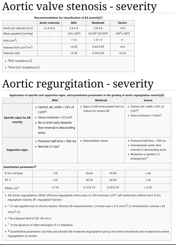
Effective regurgitant orifice (ERO) and proximal isovelocity surface area (PISA) used on echo to evaluate, dependent on volume status with ERO >0.4 cm2 used as criteria

Indications and Timing of Surgery

Performed in isolation or at time of left sided valve surgery

In Isolation

-Severe TR (ERO>0.4CM2) and isolated TR a/w excess m&m

-recent guidelines (class IIa) rec’d Tricuspid repair on its own when patients are symptomatic, including congestive hepatopathy, preferably before significant RV dysfx

-performing isolated Tricuspid repair AFTER left sided heart surgery has typically waited until severe symptoms arise leading to high M&M, however when done before severe symptoms and before significant RV dysfx outcomes are reasonable

-Severe Pulm HTN or significant RV dysfx is considered relative contraindication to surgery


TR Surgery at time of Left-sided Valve Surgery

-usually good idea because: TR doesn’t predictably improve after left sided tx, it’s not that risky to add repair of tricuspid to left repair, and reops for isolated Tricuspid repair after left repair is risky

-Risk factors for progression of TR: tricuspid annulus dilatation: ERO >40mm or 21mm/m2 on TTE or >70mm i/o; sig RV dyfx/dilatation, sig tricuspid leaflet tethering, afib, pulm htn, hx of rheumatic mitral disease, hx of RH failure

-rec’d for mild/mod TR at time of left side surgery + Tricuspid annular dilatation or RH failure


Outcomes of coronary artery bypass graft surgery

http://www.ncbi.nlm.nih.gov/pmc/articles/PMC1994021/#__abstractid201527title

This review article summarizes the major studies that have investigated the outcomes of coronary artery bypass graft surgery (CABG). The article includes a review of the literature in the areas of: history of CABG; indications for CABG; and measurement of quality of life following CABG

But it should be noted…

(recovery from cabg) is a multidimensional phenomenon that is not fully explained by medical factors. Therefore, in addition to studying mortality and morbidity outcomes following CABG, many recent studies have identified that it is important to investigate various physical, psychological, and social variables that have a significant impact on post-operative adjustment to CABG.