Pacemakers and AICDs

Keep the beat


Pacemakers and AICDs

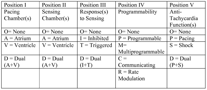
Pacemakers – trigger heartbeats based on a set rate, some can sense, some can inhibit. Some are dual inhiit and trigger.
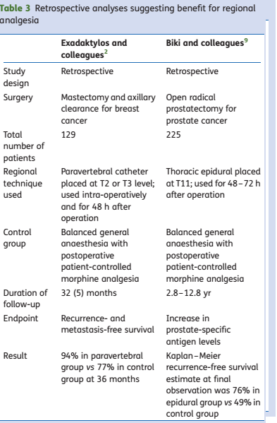
Position 1 is where they initiate
Position 2 is where they sense

ICDs – always watching R-R interval, so as to keep an eye on vtach. Either does anti-tachy pacing or shock. Most shocks are from SVT, most ICDs also have antibrady pacing functions.
Think what can be the problems

1. pacemaker gets destroyed
2. it fires when it shouldn’t
3. fails to fire when it should

For example, if it detects bovie as a beat then it might not fire when patient is actually brady. R on T is another problem that can happen. Biventricular pacing can lengthen qt interval, so defib pads essential.

Synchronous vs. Asynchronous

Asynchronous = no sensing, so allow for competitive rythm generation, so electrosurgical stim won’t interfere

3 asynchronous modes
-AOO
-VOO
-DOO

Modern ones not done like this, but good if see one that they won’t have a problem with surgical stim.
Practice Advisory
-if electromagnetic interferencelikely then set to asynchronous mode
-avoid placing magnet on an icd
-pad and bovie should not pass through pulse generator or leads
-use short/irregular lowest energy cautery
-use bipolar or harmonic
-Litho: avoid beam near pulse generator, disable atrial pacing to avoid RonT
-emergency defib: remove magnet ot reenable anti-tachycardia function

Post Op – interrogate and restore
Magnets
-use with extreme caution, you can end up turning off an ICD removing tachydysthr. detection
-most will convert to high rate asynchronous mode (80-100bpm) and less will go to asynchronous mode at a programmed rate or 60-100bpm
-removing magnet –> pacemaker mediated tachycardia from retrograde P waves picked up by dual sensing pacemakers (DDD, VDD), correct by reapplying and removing magnet
Pacemaking Modes

pacermodes

Common modes

DDD
Four rhythms possible
-NSR, Atrial sensed Ventricle paced, atrial paced ventricle sense, atrial + vent pacing
makes sure atrial event –> vent contraction
stops itself if it senses QRS
guarentees atrial kick

VVI
single chamber demand
paces entricle at preset rate(LRI is lower rate interval)
if it senses a ventricular beat it resets clock
prevents RonT
it allows for AV synchrony if atria is passing beat onto ventricle
this pacemaer can lead to pacemaker syndrome if there is loss of AV synchrony and it’s paced ventricle without any atria
Pacemaker Syndrome = fatigue, palpitaions, cough, chest fullness, cannon a waves, elevated venous pressure) and decreased CO… 25% incidence in VVI patients, severe in 5%

Other pacemaker modes

VDD – partially synchronous, pts in NSR but bad AV node. senses atria and triggers ventricle if needed but inhibits if QRS detected, also triggers when pt brady, but cannot cause atria to beat.

DDI – partially synchronous – paces atrium and ventricle at some predetermined rate but can inhibit either based on detection.

VOO – Asynchronous – fixed rate wtih no regard to pt rythm. CAN give RonT
What to do?
Pacemaker Algorithm

1. determine why pt has device, and underlying rythm
2. get a magnet, pads, atropine and isoproteronol
3. interrogate and dtermine voltage / impedence, if >2.6v or 30000, may need to replace before surgery if possible. If ICD has charge time >12s may need battery.
4. Turn off all ICD functions and use pads with EKG
5. Turn off all rate enhacements
6. Turn off minute vent response
7. Consider increasing pacing rate
8. determine magnet function
9. put defib pads on all ICD patients
10. I/O use a-line, ground placement, bipolar cautery, and disable artifact filter on ekg

TrueLearn review of the ACC/AHA Guidelines

ACC Guidelines Quick Summary

2014 ACC/AHA guidelines for non-cardiac surgery notes from TrueLearn

Cardiac event
-MI w/o intervention wait 60 days
-bare metal stent wait 30 days
-drug stent 12 months or 180 days at soonest
-14 days after balloon angio

heart failure
-CHF is higher risk
-signs/symptoms of decompensated heart failure worse

valvular disease
-moderate or greate valvular stenosis or regurg should get an echo within past year, or if any change in clinical status

arrythmias and conduction disorders
-no recs

Pulmonary vascular disease
-pulm htn adds risk
-continue with chronic pulm htn therapy

Adult congenital heart disease
-increases risk, refere to seperate guidelines

Significant arrythmias defined as (high grade av block, mobitz type 2, av block, 3rd degree av block, symptompatic vent arrythmia, Supraventricular arrythmia with uncontrolled ventricular rate, symptomatic brady, new ventricular tachycardia)

Urgency and Risk
-emergency (life/limb threatened) if not in OR within 6 hrs
-Urgent is same as above, but up to 24hrs
-Time-sensitive = delay of 1-6wks can affect outcome (oncology)
-elective = can delay up to 1 yr
-low risk = surgical and patient characteristics predict MACE <1%

The Breech

One method of handling a breech baby

Anesthesia for Breech Presentation, notes from Chestnut

Breech – when fetal buttocks/lower extremities overlie pelvic inlet

Epidemiology
-25% incidence before 28wks
-3-4% remain in breech at term


Predisposing factors
-hydrocephalus, polyhydramnios, anencephaly, oligohydramnios
-multiaprity
-hypothyroid (even transient at 12wks)
-abnormal pelvis/uterus
-previa


OB complications
-vaginal breech is high risk for neonatal trauma, c/s less so
-16x intrapartum fetal death, 3.8x intrapartum asphyxia
-5-20x cord prolapse
-13x birth trauma
-5-8% increased risk of arrest of aftercoming head**
-1/5 get spinal cord injuries with deflexion
-6-18% congenital abnormalities
-1/8 to 1/3 preterm
-5% hyperextended head

Umbilical cord prolapse necessitates emergency c/s, highest risk with incomplete breech


OB Management

External cephalic version: best done after 36-37weeks
-58% success rate, varies widely
-chance for emergent delivery
-most women who get version undergoe vaginal delivery
-some tocolytics used (Terbutaline, Nitroglycerin)
-Epidural/spinal – reduces force and pain required for version, use an anesthetic dose

Mode of delivery: c/s (Term Breech Trial 2000 http://www.ncbi.nlm.nih.gov/pubmed/11052579) preferred
-in c/s lower perinatal and neotal morbidity and mortality (1.6% vs. 5%)
-though maternal outcomes at 2yrs similiar and some experienced vaginal breech deliverers as good
-C/S can be vertical incision before 32wks
Anesthetic management

-epidural or cse good choices
-patient must not push before fully dilated as increase change for fetal head entrapment, and breech gives earlier rectal pressure, so local with opioid (fent, sufent) to block sacral segments is good as will not fully block motor in second stage
-be prepared for general at any time
-cord compression common, use supplemental O2
-if going for forceps/operative vaginal delivery, convert analgesia epidural to anasthesia with 3% chloroprocain or 2% lidocaine +/- epi and bicarb
Fetal head entrapment!!
-greatest risk less than 32wks
-head trapped in cervix 3 options:

1. duhrssen incision of cervix
-radical incisions in cervix
-maternal morbidity (trauma, hemorrhage, peritoneal bleeding)
2. relax skeletal and cervical muscle
-RSI with 2-3 MAC of halogenated agent gets relaxation in 2-3 minutes
-once delivered, siwth to 1/2mac+nitrous as high MAC ==> uterine atony/hemorrhage
-More modern approach: Nitroglycerin IV 100mcg to 200mcg, sublingual 400-800mcg Watch for hypotension and HA, tx with neo
3. c/s
-spinal, epi or GA
-increase halogenated agent in GA for relaxation
-if Neuraxial, use IV/Sublinguqal nitroglycerin
-terbutaline can also provide relaxation
-convert to GA if needed

Anesthesia and Cancer Recurrence

Notes from Br. J. Anaesth. (2010) 105 (2): 106-115. doi: 10.1093/bja/aeq164

Key points

•Metastatic disease is the most important cause of cancer-related death in patients after cancer surgery.
•Drugs and techniques used perioperatively may influence outcome.
•In vitro and animal study evidence suggests potential mechanisms altered by anaesthetic drugs.
•Human studies are limited but regional anaesthesia may be beneficial.
•There is a need for large-scale prospective studies.

Four potential mechanisms that may promote metastasis after surgery have been proposed (Table 1).

i.Handling and disrupting the tumour during surgery releases tumour cells into the circulation. Polymerase chain reaction can detect tumour cells in patient blood, and their number has been shown to increase after surgery.18
ii.The presence of the primary tumour may itself inhibit angiogenesis, and therefore, tumour removal may eliminate a safeguard against angiogenesis. This may promote survival and growth of minimal residual disease.
iii.Local and systemic release of growth factors during surgery may promote tumour recurrence both locally and at distant sites. EGF and transforming growth factor-β levels are increased, as is VEGF. In addition, anti-angiogenic factors, such as angiostatin and endostatin, may be reduced by surgery.9
iv.There is perioperative immunosuppression, including the cellular immune system. This is a result of both the neuroendocrine and cytokine stress response to surgery,19 and the effect of anaesthetic technique and other perioperative factors.


Table2_Anesthetics and Cancer

thestudies_anest_cancer


Omphalocele and Gastroschesis

total_recall_stomach_creature2
A case of unrepaired Omphalocele


From Lange: Handbook of Pediatric Anesthesia and Lange: The Anesthesia Guide

Omphalocele: viscera herniate through umbilicus, covered in peritoneum
etiology is failure of gut migration from yolk sac into abdomen
– 1:6-10k
– 2/3 have congenitial abnormalities: CV(significantly increasese mortality) , GU(bladder extrophy), GI (meckel’s, malrotation), Cranio, trisomy 13, Bckwith-Widemann (visceromegaly, macroglossia, microcephaly, hypoglycemia)
-survival rate: 70-95%
Gastroschisis: no covering, defect lateral to umbilicus (right)
etiology is occlusion of omphalomesenteric artery with ischemia to the right of periumbilical area
– 1:30k
– low incidence of associated abnormalities, but if so similiar to omphalocele; GI intestinal atresia
-premature labor and devliery common, more issues with bowel function
-survival rate: >90%

Both dx prenatal u/s
Both  M>F
Both a/w Trisomy 21, congential diaphragmatic hernia
Pre-Op

NG tube to decompress stomach
Broad-spectrum A/B
Expedited surgery to minimze heat loss, infection, trauma
Fluid resuc. 150-300mg/kg/d with balanced salt soln., maintain 1-2ml/kg/h UOP
urgent surgery, but w/u associated abnormalities: echo, renal u/s
Anesthetic Management

-suction stomach and pre-oxygenate
-awake or RSI with ETT
-fluid resucitation
-prevent hypothermia
-opioid + muscle relaxant (nondep)
-pulse ox on lower extremity can detect congestion d/t obstructed venous return
+/- a line (esp. if cardiac)
– increased intra-abdominal pressure warnings: vent compromise (peak airway pressure higher, decreased TV), bowel edema, anuria, hypotension, decreased organ perfusion)
*if insp pressure >25-30 or intragastric >20, don’t do primary closure

P/O
-ventilation for 24-48 hrs, monitor airway pressure, fluid resuc., a/b
-watch out: abdominal compartment syndrome (gastric pressure<20, vesical <20,  peak vent pressure<30cm/h20)

Do’s and Don’ts
-Do: pre-op echo for omphalocele
-Do: aggressive fluid resuc.
-Do: communicate to surgeon about tight closure
-Do: adequated nondepolarizer for closure
-DON’T: Mask ventilate!
Surgical concerns
-small defect: primary closure
-large defect: staged closure with silastic silo which is secured at edge and reduced over about a week, when pt goes to OR for closure